TNF Counterbalances the Emergence of M2 Tumor Macrophages. One-way ANOVA indicated a significant difference in PD-1 mRNA levels between the In these studies, Lyz2hiF4/80+CD11b+ mature tissue macrophages have been identified as the critical M(IL-4) population suppressing inflammation in the liver, while LyzloF4/80+CD11b+ monocytes expressing high amounts of arginase-1 were largely responsible for the inhibition of fibrosis during chronic infection. Macrophage necroptosis, a form of programmed necrosis characterized by the death of inflammatory cells has been recently identified as a key signal maintaining microbial induced type 1 inflammation.
Phenotypic and functional plasticity of cells of innate immunity: macrophages, mast cells and neutrophils. Deng P, Qiu S, Liao F, Jiang Y, Zheng C, Zhu Q. Exp Biol Med (Maywood). official website and that any information you provide is encrypted Mmp12 activity is also increased in CCL4 and thioacetamide-induced liver fibrosis, but in these models Mmp12 activity had either no effect or led to slightly decreased fibrosis (Pellicoro et al., 2012). CCL2-dependent infiltrating macrophages promote angiogenesis in progressive liver fibrosis. Using several elegant in vivo strategies to globally deplete monocytes and macrophages, Gibbons and colleagues have concluded that macrophages are critically required for the development of bleomycin induced pulmonary fibrosis (Gibbons et al., 2011).
 Bourdonnay and colleagues have also identified trans-cellular delivery of vesicular suppressor of cytokine signaling (SOCS) proteins as another unique form of intercommunication between AMs and epithelial cells, a mechanism that also plays important roles in the resolution of inflammation in the lung (Bourdonnay et al., 2015). WebFibrosis involves scar formation and normal function is not restored. Macrophages decide between regeneration and fibrosis in muscle. Boulter L, Govaere O, Bird TG, Radulescu S, Ramachandran P, Pellicoro A, Ridgway RA, Seo SS, Spee B, Van Rooijen N, et al. Irf5 deficiency in macrophages promotes beneficial adipose tissue expansion and insulin sensitivity during obesity. Tissue repair and regeneration are critical biological processes that are fundamental to the survival of all living organisms (Das et al., 2015). Local arginase 1 activity is required for cutaneous wound healing. WebRegeneration vs Fibrosis. Thus, VEGF- production by macrophages was identified as an indispensable mechanism in nerve regeneration. These studies are important because they suggest functionally distinct CD11b+ macrophages regulate the injury and recovery phases of tissue repair (Duffield et al., 2005). Inflammatory responses that develop following tissue injury are associated with the production of a variety of inflammatory mediators like IFN- and TNF- that promote classical macrophage activation. However, the contribution of macrophages to the development and maintenance of IL-13-dependent fibrosis is less clear as macrophages are not thought to be a major source of IL-13 (Wynn, 2004). Jenkins SJ, Ruckerl D, Cook PC, Jones LH, Finkelman FD, van Rooijen N, MacDonald AS, Allen JE. Regeneration and fibrosis share a common cascade of injury-induced events that bifurcates as a result of the chronicity of the damage . The https:// ensures that you are connecting to the Fibrosis is the first step in the regeneration process. Using a radiation chimera model to distinguish bone marrow-derived cells from microglia, Evans and colleagues have determined that the vast majority of accumulated cells in spinal cord injury are derived from the blood, and CX3CR1+ macrophages but not CCR2+ monocytes were tightly associated with axonal dieback (Evans et al., 2014). Selective depletion of macrophages reveals distinct, opposing roles during liver injury and repair. Yun MH, Davaapil H, Brockes JP. It occurs in tissues that are capable of completely rebuilding Recent studies have suggested that yolk sac-derived resident tissue macrophages and monocytes recruited from the bone marrow play distinct roles during the different stages of repair in some organs. arrow_forward. They have shown that AMs secrete SOCS1 and -3 exosomes and microparticles, respectively, which are taken up by alveolar epithelial cells leading to the suppression of Stat activation (Figure 3). Before Shouval DS, Biswas A, Goettel JA, McCann K, Conaway E, Redhu NS, Mascanfroni ID, Al Adham Z, Lavoie S, Ibourk M, et al. Following CNS demyelination, microglia and peripherally derived inflammatory macrophages switch from a pro-inflammatory or classically activated M(IFN-) phenotype to a pro-repair or alternatively-activated M(IL-4)-like phenotype as repair commences, and intra-lesional M(IL-4) cell depletion substantially delayed oligodendrocyte differentiation (Miron et al., 2013). They have shown that development of early onset inflammatory bowel disease (IBD) in IL-10R-deficient patients is also associated with major defects in the generation and function of anti-inflammatory macrophages. Qin H, Luo Z, Sun Y, He Z, Qi B, Chen Y, Wang J, Li C, Lin W, Han Z, Zhu Y. Int J Biol Sci. Indeed, if macrophages are depleted early after injury, the inflammatory response is often greatly diminished (Duffield et al., 2005). and transmitted securely. Regenerative macrophages also facilitate skeletal muscle regeneration by directly targeting myogenic precursor cells (MPCs). Inflammatory monocytes and resident tissue macrophages are key regulators of tissue repair, regeneration, and fibrosis. Thus, macrophages exhibiting a pro-fibrotic phenotype participate in the activation and expansion of ECM-producing myofibroblasts via multiple mechanisms.
 Distinct pro-inflammatory and wound healing macrophage phenotypes have also been observed in models of spinal cord injury and repair, with the functionally distinct macrophage populations recruited to the site of tissue injury by unique chemokine gradients. WebTissues are repaired by fibrosis and regeneration. Wehr A, Baeck C, Ulmer F, Gassler N, Hittatiya K, Luedde T, Neumann UP, Trautwein C, Tacke F. Pharmacological inhibition of the chemokine CXCL16 diminishes liver macrophage infiltration and steatohepatitis in chronic hepatic injury. For example, Chen and colleagues have shown that CCL2 is released from neighboring hair follicles when hairs are plucked, leading to the rapid recruitment of TNF- secreting inflammatory macrophages, which accumulate near the plucked hair and provide key signals to local stem cells that facilitate the regeneration of new hair follicles (Chen et al., 2015a). Stutchfield and colleagues also have shown that CSF1-Fc therapy could be used to expand the numbers of protective tissue macrophages in models of acute liver injury and partial hepatectomy (Stutchfield et al., 2015). Pathophysiology: The body has a remarkable way of healing, specifically with tissue repair.
judith goss parcells Pulmonary macrophage transplantation therapy. The site is secure. Thus, nutrient competition between local tissue macrophages and neighboring immune cells has been identified as an additional potent immunosuppressive mechanism employed by regulatory macrophages (Murray et al., 2015). TGF-beta driven lung fibrosis is macrophage dependent and blocked by Serum amyloid P. Murray PJ, Allen JE, Biswas SK, Fisher EA, Gilroy DW, Goerdt S, Gordon S, Hamilton JA, Ivashkiv LB, Lawrence T, et al. Therefore it is difficult to conclude definitively whether fibrosis is dependent on M(IL-4) cells, although adoptive transfer studies suggest this was likely the case. They determined that pro-fibrotic macrophage function is highly dependent on TNF- and IL-1-induced survival but not activation of hepatic stellate cells in vitro and in vivo. Regeneration: Regeneration is a key biological process that is essential to an organism's life. Mitchell S, Thomas G, Harvey K, Cottell D, Reville K, Berlasconi G, Petasis NA, Erwig L, Rees AJ, Savill J, et al. Sustained IL-1 production through NLRP3 inflammasome activation in macrophages has also been shown to be a major driver of persistent inflammation and fibrosis in other tissues as well, including the liver during chronic hepatitis C virus infection (Negash et al., 2013). Give examples of the cells and tissues involved in both repair processes. M(IL-4) cells, in turn, establish an anti-inflammatory environment that is more accommodating to the survival and growth of both mesenchymal stem cells and progenitor populations in injured tissues, suggesting a mutually beneficial feed-back loop exists between anti-inflammatory macrophages and stem cell populations that drive tissue regeneration (Freytes et al., 2013; Mounier et al., 2013). 18 20 This provides a richness of potential therapeutic targets to reduce fibrosis and facilitate skeletal muscle regeneration. Lech M, Anders HJ. Fibrosis is typically associated with impaired angiogenesis and sustained development of local tissue hypoxia, with hypoxia-inducible factor-1a (HIF-1a), a transcription factor that functions as a master regulator of oxygen homeostasis directly implicated in TGF-1-driven fibrogenesis (Ueno et al., 2011).
Consequently, CSF-1 protein, antibodies against the ligand and receptor, and inhibitors of CSF-1R kinase activity are all being tested in various disease models and more recently clinical trials have also been initiated. An official website of the United States government. Xu J, Chi F, Guo T, Punj V, Lee WN, French SW, Tsukamoto H. NOTCH reprograms mitochondrial metabolism for proinflammatory macrophage activation. Type 2 innate signals stimulate fibro/adipogenic progenitors to facilitate muscle regeneration. A similar role for IL-10R signaling in macrophages has been recently generated in a model of corneal lymphangiogenesis. With repeated exposure to the injurious agent, HHS Vulnerability Disclosure, Help Macrophages are critical to the maintenance of IL-13-dependent lung inflammation and fibrosis. These authors have found that the route of monocyte and macrophage entry to the central nervous system also provides additional instructional signals to shape the unique functional activities of the recruited cells. Jenkins SJ, Ruckerl D, Thomas GD, Hewitson JP, Duncan S, Brombacher F, Maizels RM, Hume DA, Allen JE.
 Consequently, because they represent potentially important therapeutic targets, there has been great deal of interest over the past few years in deciphering the contributions of the different macrophage populations that control the initiation, maintenance, and resolution of wound healing responses in different organ systems. WebRegeneration is healing taken to the next level. The resident tissue macrophages are thought to quickly convert to a pro-resolution tissue repair phenotype during AALF, so expanding their numbers through local proliferation or recruitment from the monocyte pool has been hypothesized to be a critical determinant controlling survival following severe liver injury. Thus, a variety of mechanisms, besides IL-10, are involved in the development of macrophages with pro-wound healing and anti-inflammatory activity. This review focuses on recent findings that have advanced our understanding of the role of monocytes and resident tissue macrophages in wound repair, tissue regeneration, and fibrosis. Two recent papers have addressed this question in models of mucosal healing by performing adoptive transfer studies and generating mice with genetic deletions of IL-10 or the IL-10 receptor alpha chain in macrophages. Purpose of review: In support of this conclusion, recent studies of AALF and partial hepatectomy in mice showed that by expanding the number of resident tissue macrophages and recruited monocytes exhibiting a reparative anti-inflammatory phenotype, colony-stimulating factor 1-Fc treatment holds promise as a therapeutic strategy following acute liver injury (Stutchfield et al., 2015). They also demonstrate how the timely conversion of monocytes and macrophages from a pro-inflammatory to a reparative phenotype plays a decisive role in wound healing and tissue regenerative responses.
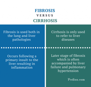
 The main causes of liver fibrosis include chronic viral infection, alcohol abuse, and nonalcoholic steatohepatitis, while pulmonary fibrosis is caused by a family of more than 200 chronic lung diseases including scleroderma (systemic sclerosis), sarcoidosis, infections, exposure to toxicants or radiation, and idiopathic pulmonary fibrosis. Wynn TA, Barron L. Macrophages: master regulators of inflammation and fibrosis. Finally, N-3 polyunsaturated fatty acids, which exhibit substantial anti-inflammatory in vivo, confer important cardio protective effects by inhibiting macrophage-mediated activation of cardiac fibroblasts. The manuscript will undergo copyediting, typesetting, and review of the resulting proof before it is published in its final citable form. Macrophage production of transforming growth factor beta and fibroblast collagen synthesis in chronic pulmonary inflammation.
Organ-level quorum sensing directs regeneration in hair stem cell populations. Jay TR, Miller CM, Cheng PJ, Graham LC, Bemiller S, Broihier ML, Xu G, Margevicius D, Karlo JC, Sousa GL, et al. A new, and perhaps surprising, relationship between fibrosis regression and angiogenesis is revealed by Kantari-Mimoun et al. Fibrotic disease and the T(H)1/T(H)2 paradigm. Fibrosis may just be a functionally irrelevant replacement of damaged tissue or even help to preserve structural integrity of the remaining tissue. In addition, chronic hepatitis often accompanies proliferation of atypical biliary cells, also known as liver progenitor cells or oval cells. In addition to communicating anti-inflammatory signals directly to epithelial cells, macrophages also regulate communication in the epidermis. Tissue repair occurs by two methods: fibrosis and regeneration.Fibrosis is the formati View the full answer Previous question Next question Similar studies conducted in other tissues identified regenerating islet-derived 3 beta (Reg3) as an essential regulator of macrophage trafficking to cardiac tissues following injury (Lorchner et al., 2015). eCollection 2023. Wynn TA, Ramalingam TR. Madala SK, Pesce JT, Ramalingam TR, Wilson MS, Minnicozzi S, Cheever AW, Thompson RW, Mentink-Kane MM, Wynn TA. They are regenerative phase and fibrosis. In the regenerative phase, the injured cells are replaced by the same kind of cells. In fibrosis, connective tissue replaces normal parenchyma tissue. 1. Overview and Key Difference 2. What is Regeneration Epub 2022 Jul 1. It is the proliferative capacity of the cells that decides the amou. Shechter R, Miller O, Yovel G, Rosenzweig N, London A, Ruckh J, Kim KW, Klein E, Kalchenko V, Bendel P, et al. The distinct tissue macrophage populations that take up residence in many tissues of the body are mostly derived from the yolk sac during embryogenesis, with fetal liver and hematopoietic stem cells contributing macrophages to some but not all tissues at later time points (Epelman et al., 2014a; Epelman et al., 2014b; Gomez Perdiguero et al., 2015). Consequently, they have hypothesized it might have little impact on the maintenance of inflammatory disease. Fibrosis noun. The multi-cellular process is initiated by injury induced hypoxia, which is sensed by local tissue macrophages that then secrete VEGF- to induce a polarized vasculature that relieves the hypoxia, but at the same time it creates a path for proliferating Schwann cells to migrate across to reconnect the nerve. Pellicoro A, Aucott RL, Ramachandran P, Robson AJ, Fallowfield JA, Snowdon VK, Hartland SN, Vernon M, Duffield JS, Benyon RC, et al. kibana hardware requirements; adam carlyle taylor obituary; difference between fibrosis and regeneration; by in pigeon meat for bell's palsy. Inflammatory monocytes and resident tissue macrophages are key regulators of tissue repair, regeneration, and fibrosis. Regeneration is the idea that the body can regrow parts of itself after an injury. While regeneration describes the specific substitution of the tissue, i.e. Wound macrophages express TGF-alpha and other growth factors in vivo: analysis by mRNA phenotyping. Interleukin-4 Receptor alpha Signaling in Myeloid Cells Controls Collagen Fibril Assembly in Skin Repair.
Thus, recruited bone marrow derived monocytes exhibit tissue destructive activity while embryonic-derived resident tissue populations facilitate the resolution of inflammation and instruct tissue repair in the heart. It also remains unclear whether an individual macrophage (local or recruited) is capable of adopting all of these attributes at different times in response to signals found in the local tissue microenvironment or whether there are truly distinct functional subsets of monocytes and macrophages that are hard-wired to regulate these different and often opposing activities. Figure 1 Open in figure viewer PowerPoint Likewise, although there are obvious parallels between fibrosis in the kidney and elsewhere, there are also a number of important differences, and kidney specific consequences, that distinguish progressive renal disease. WebFibrosis, regeneration and cancer: what is the link?
(medicine) The formation of (excess) fibrous connective tissue in an organ.
 These studies are of interest because they suggest unique roles for different populations of IL-4 and/or IL-13-activated inflammatory monocytes and resident tissue macrophages in the resolution of inflammation, tissue repair, and fibrosis. WebRegeneration. Zhang MZ, Yao B, Yang S, Jiang L, Wang S, Fan X, Yin H, Wong K, Miyazawa T, Chen J, et al. Damage associated molecular patterns (DAMPs), PAMPs (pathogen associated molecular patterns), Regulatory T cells (Treg), interferon-regulatory factor 5 (IRF5), nitric oxide synthase 2 (NOS2), Liver X receptor (LXR), Amphiregulin (AREG), Arginase-1 (Arg1), interferon regulatory factor 4 (IRF4), peroxisome proliferator-activated receptor gamma (PPAR), fibroblast growth factor (FGF), galectin-3 (GAL-3), transforming growth factor (TGF), Immune complex (IC), glucocorticoid receptor (GR), transcription factor ATF3, silencers of cytokine signaling (SOCS). 8600 Rockville Pike Alveolar macrophages transmit key signals to neighboring cells that help facilitate the resolution of inflammation in the lung. WebWhat is the difference between fibrosis and regeneration? Macrophage activation and polarization: nomenclature and experimental guidelines. Nephrol Dial Transplant. PMC Please note that during the production process errors may be discovered which could affect the content, and all legal disclaimers that apply to the journal pertain. Ramachandran P, Pellicoro A, Vernon MA, Boulter L, Aucott RL, Ali A, Hartland SN, Snowdon VK, Cappon A, Gordon-Walker TT, et al. As a service to our customers we are providing this early version of the manuscript. The IL-21 receptor augments Th2 effector function and alternative macrophage activation. 2005 Dec;33(12):1816-24. doi: 10.1177/0363546505278701. Multi-potent adult progenitor cells have also been shown to exhibit similar M(IL-4) polarizing activity, leading to a reduction in macrophage mediated axonal dieback in spinal cord injury (Busch et al., 2011). Regeneration = tissue repaired to normal state Healing = Repair occurs by lying down connective tissue; scar CICATRIZATION: Substitution of the injured tissue by connective tissue stroma (scar). Macrophage activation and skeletal muscle healing following traumatic injury. Interleukin-10 receptor signaling in innate immune cells regulates mucosal immune tolerance and anti-inflammatory macrophage function. Suzuki T, Arumugam P, Sakagami T, Lachmann N, Chalk C, Sallese A, Abe S, Trapnell C, Carey B, Moritz T, et al. Chen F, Liu Z, Wu W, Rozo C, Bowdridge S, Millman A, Van Rooijen N, Urban JF, Jr, Wynn TA, Gause WC. What is meant by tissue regeneration? Epelman S, Lavine KJ, Randolph GJ. It also regulates their migration, proliferation, function, and survival. Miron VE, Boyd A, Zhao JW, Yuen TJ, Ruckh JM, Shadrach JL, van Wijngaarden P, Wagers AJ, Williams A, Franklin RJ, ffrench-Constant C. M2 microglia and macrophages drive oligodendrocyte differentiation during CNS remyelination. Factors that prevent accumulating tissue monocytes from converting from a pro-inflammatory to reparative phenotype can also impair healing.
 The main difference between the two processes is regeneration results in the damaged cells replaced by identical cells through mitosis while fibrosis replaces the damaged cells with a network of collagen and . Although approaches that either reduce the numbers of inflammatory macrophages exhibiting an M(IFN-) skew phenotype or increase the numbers of reparative anti-inflammatory M(IL-4)-like macrophages have been shown to accelerate the repair of many tissues, persistent activation or sustained recruitment of the M(IL-4)-like cells has also been hypothesized to contribute to the development of pathological fibrosis (Wynn and Ramalingam, 2012). CSF1 Restores Innate Immunity After Liver Injury in Mice and Serum Levels Indicate Outcomes of Patients With Acute Liver Failure. One relatively straightforward way to manipulate macrophage function is to regulate their numbers by targeting CSF1 and CSF-1R signaling, as CSF1 is critically required for the differentiation of myeloid progenitors into heterogeneous populations of monocytes and macrophages (Hume and MacDonald, 2012).
As illustrated in this review, monocytes and macrophages are recruited and activated by several distinct mechanisms and assume many functional characteristics that are critical to tissue injury and repair. Although pro-inflammatory and anti-inflammatory macrophages are the two most frequently investigated phenotypes in studies of wound repair, fibrosis and tissue regeneration, macrophages exhibiting pro-wound healing, pro-fibrotic, anti-fibrotic, pro-resolving, and tissue regenerating characteristics are also commonly mentioned in the literature. Wynn TA. Cells residing between the hepatocytes and small blood vessels in the liver. Finally, by mimicking the anti-inflammatory effects of apoptotic cell, phosphatidlyserine-presenting liposomes have also been used to induce reparative IL-10 and TGF1-producing cardiac macrophages (Harel-Adar et al., 2011). obtained complimentary findings that showed macrophage-derived IL-10 is dispensable for gut homeostasis and maintenance of colonic Treg cells (Zigmond et al., 2014). Finally, Knipper and colleagues have investigated a model of skin repair and showed that collagen fibril assembly following injury is also highly dependent on M(IL-4) macrophages (Knipper et al., 2015). IL-25 induces M2 macrophages and reduces renal injury in proteinuric kidney disease. Together, the preceding studies, which encompass various organ systems and experimental models, nicely illustrate the distinct and often opposing roles of inflammatory monocytes and resident tissue macrophages in tissue repair. No products in the cart. Regeneration replaces damaged tissue with scar formation and normal function is not restored. Following tissue injury, monocytes and Pradere JP, Kluwe J, De Minicis S, Jiao JJ, Gwak GY, Dapito DH, Jang MK, Guenther ND, Mederacke I, Friedman R, et al. Resident tissue macrophages and Kupffer cells have also been shown to play a key role in the liver following acetaminophen-induced acute liver failure (AALF) in mice and humans. Fibrosis is the formation of excess fibrous connective tissue in an organ or tissue Although FIBROSIS can be caused by things other than injury, Stutchfield BM, Antoine DJ, Mackinnon AC, Gow DJ, Bain CC, Hawley CA, Hughes MJ, Francis B, Wojtacha D, Man TY, et al. Pradere and colleagues showed that hepatic macrophages enhance myofibroblast survival by stimulating nuclear factor kappa B (NF-b) activity in fibroblasts, which is critical for the development of liver fibrosis (Pradere et al., 2013). Thus, in future work, it will be important to include as many distinguishing characteristics about the macrophages being studied as possible (Murray et al., 2014), including cell surface markers and gene expression analyses that reveal transcriptional and epigenetic profiles, as this will increase our ability to compare findings between research groups and expand our understanding of the unique contributions of the different macrophage populations and activation states during tissue injury and repair in multiple organ systems. Here, programmed suicide of infected Kupffer cells triggers significant monocyte recruitment and anti-microbial type 1 immunity (Bleriot et al., 2015). What are the differences between the two types of tissue repair? Zigmond E, Bernshtein B, Friedlander G, Walker CR, Yona S, Kim KW, Brenner O, Krauthgamer R, Varol C, Muller W, Jung S. Macrophage-restricted interleukin-10 receptor deficiency, but not IL-10 deficiency, causes severe spontaneous colitis. Neutrophil extracellular traps license macrophages for cytokine production in atherosclerosis. WebTissues are repaired by fibrosis and regeneration. Mitchell C, Couton D, Couty JP, Anson M, Crain AM, Bizet V, Renia L, Pol S, Mallet V, Gilgenkrantz H. Dual role of CCR2 in the constitution and the resolution of liver fibrosis in mice. Cholesterol crystals trigger neutrophils to release extracellular traps (NETs), which prime local macrophages to transcribe immature IL-1, with cholesterol crystals serving a second role as a danger signal that activates inflammasomes, which process immature IL-1 for secretion.
 Busch SA, Hamilton JA, Horn KP, Cuascut FX, Cutrone R, Lehman N, Deans RJ, Ting AE, Mays RW, Silver J. Multipotent adult progenitor cells prevent macrophage-mediated axonal dieback and promote regrowth after spinal cord injury.